< Developed Cellular Mechanics Programs >
We combine nanoscale experiments with computational models to understand how cell membranes and their cytoskeletons govern mechanics, adhesion, and molecular transport.
Red Blood Cell Mechanics and Adhesion
Experiments. We measure membrane mechanics and adhesion in healthy and sickle red blood cells. Our Biophysical Journal studies examine how AKAP-organized signaling regulates BCAM/Lu binding to laminin (2014) and the activation of ICAM-4 adhesion receptors (2017), linking intracellular signaling to interactions with vascular surfaces.
Simulations. Our coarse-grained molecular dynamics models explicitly represent the lipid bilayer, spectrin network, and membrane proteins. We investigate membrane deformation, band-3 protein diffusion, vesicle formation, and the effects of membrane defects in blood disorders.
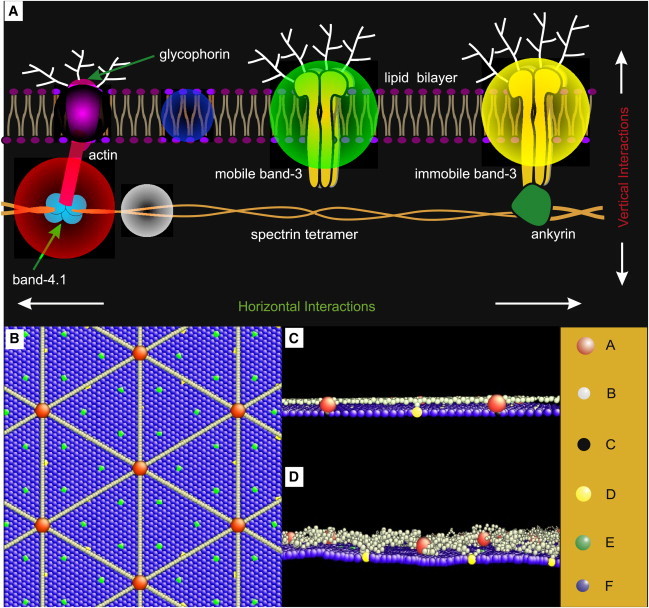

Maciaszek et al., Biophysical Journal (2014). Cover: © Biophysical Society
Selected Biophysical Journal papers: Membrane model — Li & Lykotrafitis (2014) · BCAM/Lu adhesion — Maciaszek et al. (2014) · ICAM-4 regulation — Zhang et al. (2017)
Neurons and Neuronal Axons
We combine microscopy, force measurements, single-particle tracking, and molecular simulations to study how membrane and cytoskeletal organization influence axon mechanics, ion-channel localization, and protein diffusion.
Experiments
Confocal Microscopy and Channel Localization
Confocal imaging identifies the axon initial segment and maps potassium-channel subtypes. Abiraman, Tzingounis, and Lykotrafitis showed that KCa2.3 is the predominant KCa2 subtype in this region. Dual-color labeling distinguishes surface and internalized channels, revealing compartment-specific trafficking controlled by cAMP–PKA signaling. FASEB Journal (2018), confocal images in Figs. 2–4.
Force Nanoscopy and Native SK-Channel Organization
Maciaszek and colleagues (2012) mapped native SK channels in living hippocampal neurons using force nanoscopy. Channels were enriched in distal dendrites and commonly detected as single channels or pairs. PKA signaling dynamically regulated their distribution, establishing a foundation for our later fluorescence imaging and diffusion studies. Read the Journal of Neuroscience paper
TIRF Microscopy and Protein Diffusion
Total internal reflection fluorescence (TIRF) microscopy tracks quantum-dot-labeled SK channels in living neurons. Our measurements show that actin organization, neuronal maturation, and membrane crowding regulate channel mobility differently in the soma, dendrites, and axon initial segment.
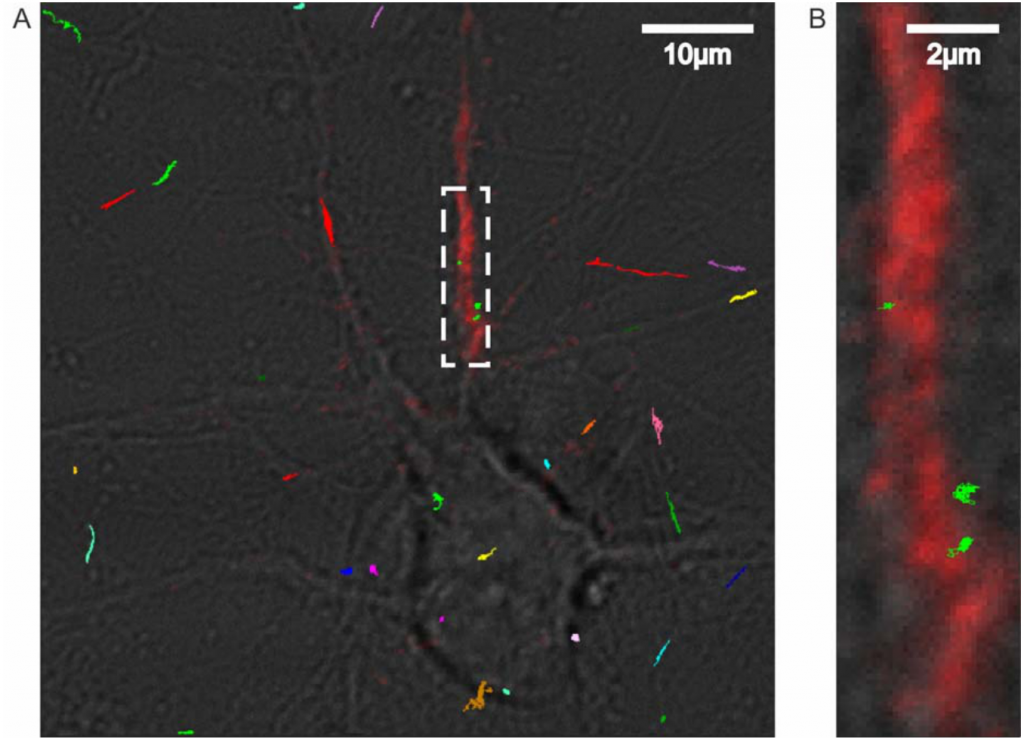
Tracking SK-channel motion. Colored trajectories overlay the neuron; the enlarged inset shows the axon initial segment.
Gu, Tzingounis & Lykotrafitis, Function (2023), Fig. 1. Author manuscript.
Simulations
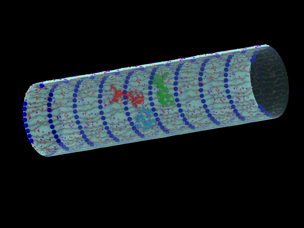
Our coarse-grained molecular dynamics models represent the periodic actin–spectrin skeleton and its coupling to the lipid bilayer and membrane proteins. We use these models to investigate axon mechanics and protein transport within and between membrane compartments.
Axon Membrane Architecture and Mechanics
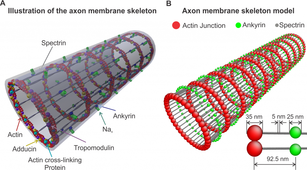
Zhang et al., PLOS Computational Biology (2017), Fig. 1. CC BY.
The model explicitly represents actin rings connected by spectrin filaments and ankyrin. This organization provides a basis for studying the mechanical response and stability of the axon membrane skeleton.
Simulations of protein diffusion
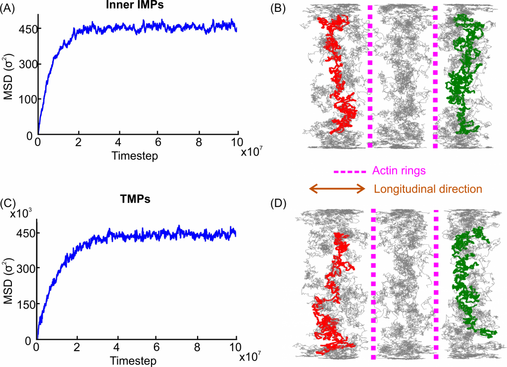
Zhang, Tzingounis & Lykotrafitis, PLOS Computational Biology (2019), Fig. 5. CC BY.
Our molecular model couples the lipid bilayer, actin–spectrin skeleton, and membrane proteins. Simulated trajectories and mean-squared displacement show how actin rings restrict longitudinal protein motion, while spectrin and protein crowding modify diffusion within membrane compartments.